Ever wondered what's actually inside the cells that make up...
Comprehensive LC Biology Notes and Diagrams
C
Chloe Ahern@chloeahern
1 / 10
1
of 10
Cell Structure Basics
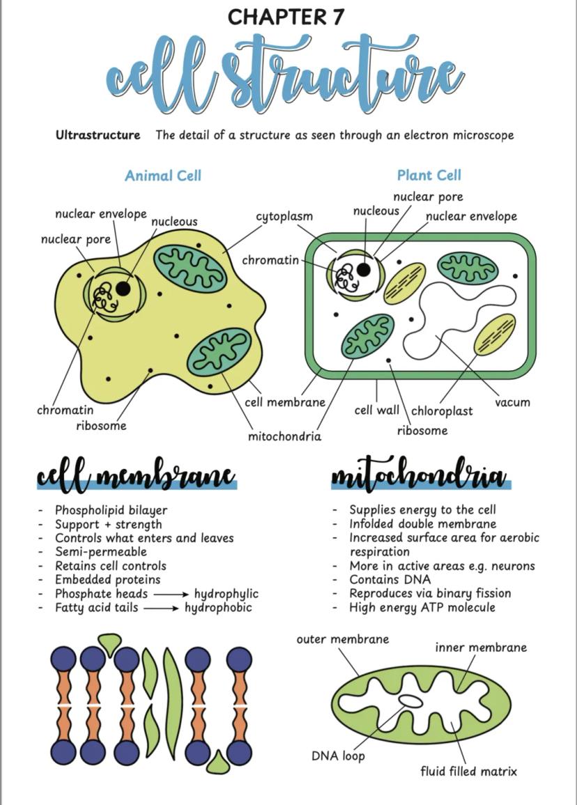
Your body is made up of trillions of cells, and understanding their ultrastructure (what you see under powerful microscopes) is key to biology success. Think of cells as tiny factories with different departments doing specific jobs.
Animal cells and plant cells share many features but have crucial differences. Both have a nucleus (the control centre), cytoplasm , and ribosomes (protein makers). The cell membrane acts like a selective bouncer, controlling what gets in and out using its phospholipid bilayer structure.
Mitochondria are your cell's power stations, producing energy through aerobic respiration. They're packed with folded membranes to maximise surface area - you'll find loads of them in active cells like neurons that need tons of energy.
Quick Tip: Remember that mitochondria have their own DNA and reproduce independently - they're like tiny organisms living inside your cells!
2
of 10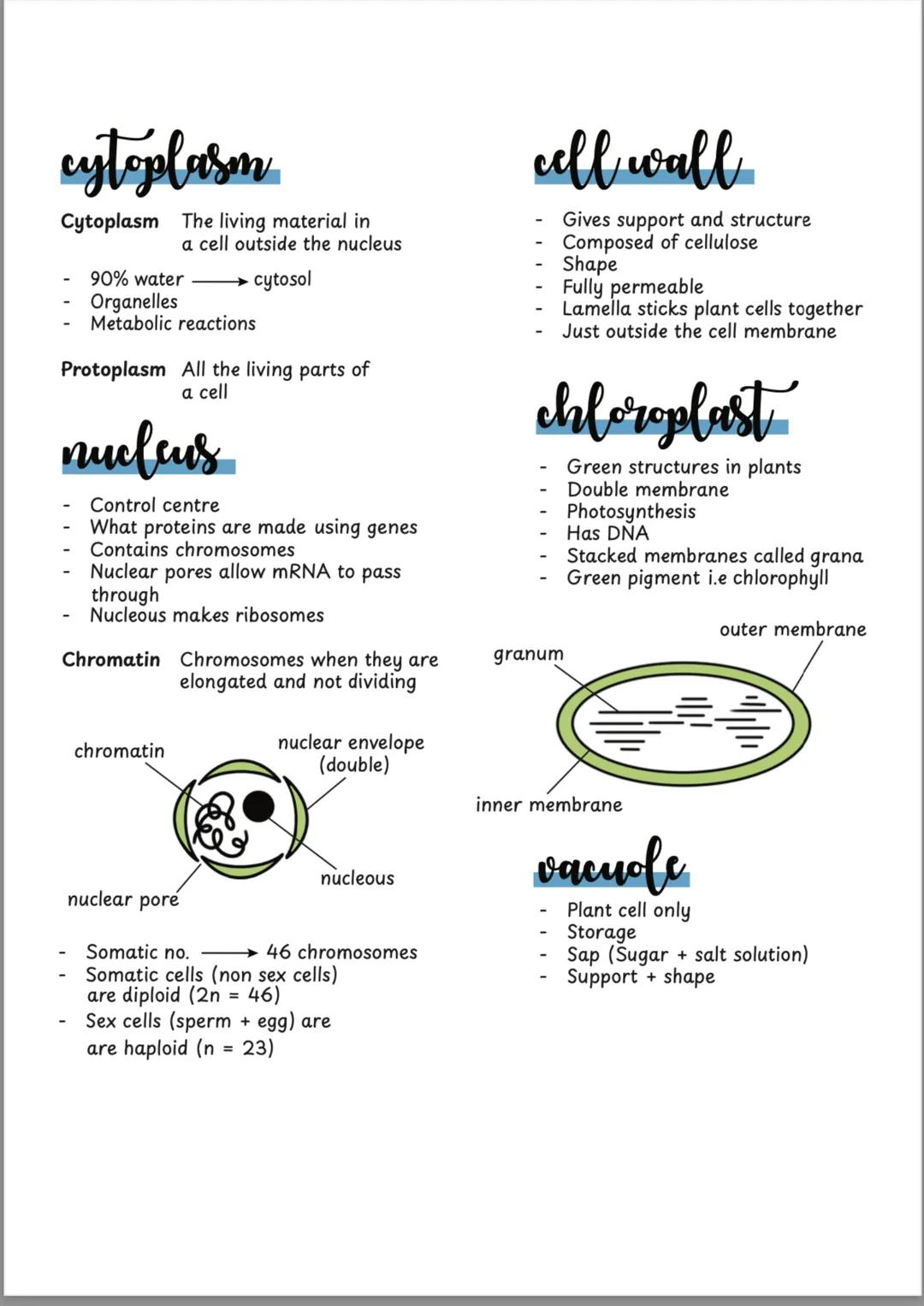
Plant vs Animal Cells
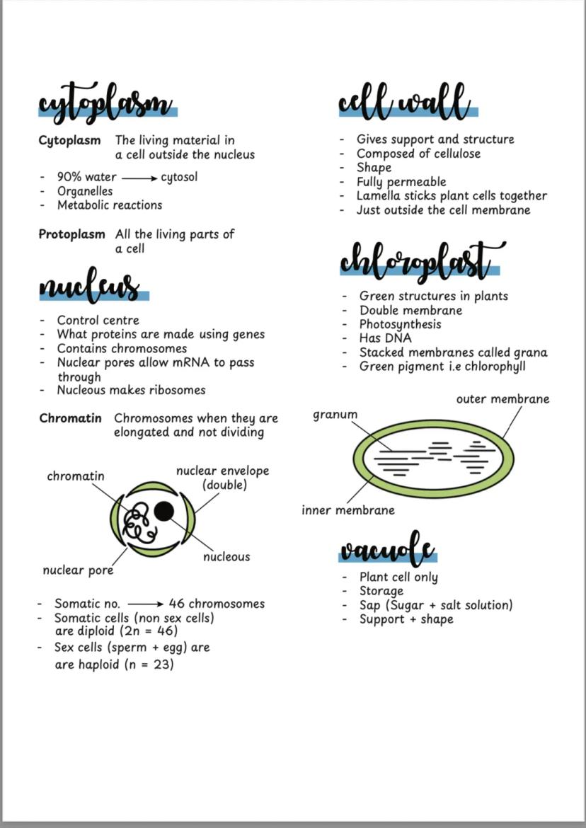
Plant cells have some extra features that animal cells don't. The cell wall made of cellulose gives plants their rigid structure and shape - imagine it as a protective outer shell around the cell membrane.
Chloroplasts are the green powerhouses where photosynthesis happens. These double-membraned organelles contain chlorophyll and have stacked membranes called grana that capture sunlight. Plant cells also have a large central vacuole filled with sap that helps maintain the plant's shape and stores materials.
The nucleus is the brain of any cell, containing chromosomes (which become chromatin when not dividing). Nuclear pores let important molecules like mRNA pass through. Cytoplasm is 90% water and houses all the organelles where metabolic reactions occur.
Remember: All the living parts of a cell together are called protoplasm - this includes everything except the cell wall!
3
of 10
Prokaryotic vs Eukaryotic Cells
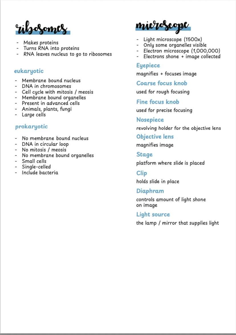
Understanding the difference between eukaryotic and prokaryotic cells is crucial for your exams. Eukaryotic cells (like yours) have a membrane-bound nucleus and organelles - they're the advanced, complex cells found in animals, plants, and fungi.
Prokaryotic cells are much simpler - think bacteria. They have no membrane-bound nucleus, just DNA floating in a circular loop. No fancy organelles either, and they're much smaller than eukaryotic cells.
Light microscopes can magnify up to 1,500 times and show basic cell structures. Electron microscopes are the real game-changers, magnifying up to 1,000,000 times and revealing incredible detail. Understanding how to use a light microscope properly is essential lab skill.
Lab Success: Always start with low power objective lens, use coarse focus first, then switch to high power and use fine focus for crisp images!
4
of 10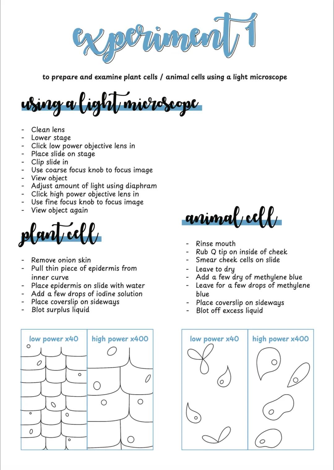
Microscope Experiments
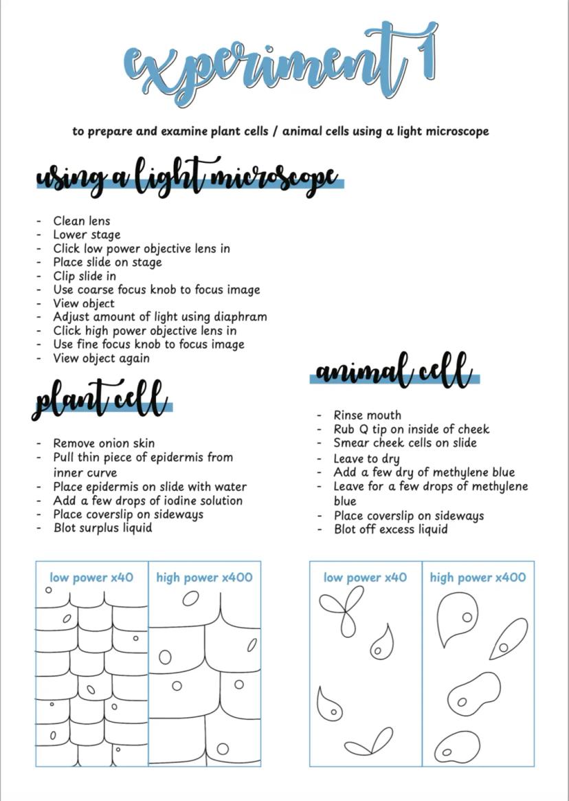
Preparing cell samples is a key practical skill you'll need to master. For plant cells, onion epidermis works brilliantly - it's thin, easy to handle, and shows cell walls clearly when stained with iodine solution.
Animal cells from your cheek are perfect for observation. A gentle rub with a cotton bud gives you plenty of cells to examine. Methylene blue stain makes the nucleus and cell membrane visible under the microscope.
The technique is crucial: place your sample on a slide with a drop of water, add stain, then lower the coverslip at an angle to avoid air bubbles. Always blot excess liquid to prevent mess and improve image quality.
Pro Tip: When switching from low to high power, always centre your specimen first - high power has a much smaller field of view!
5
of 10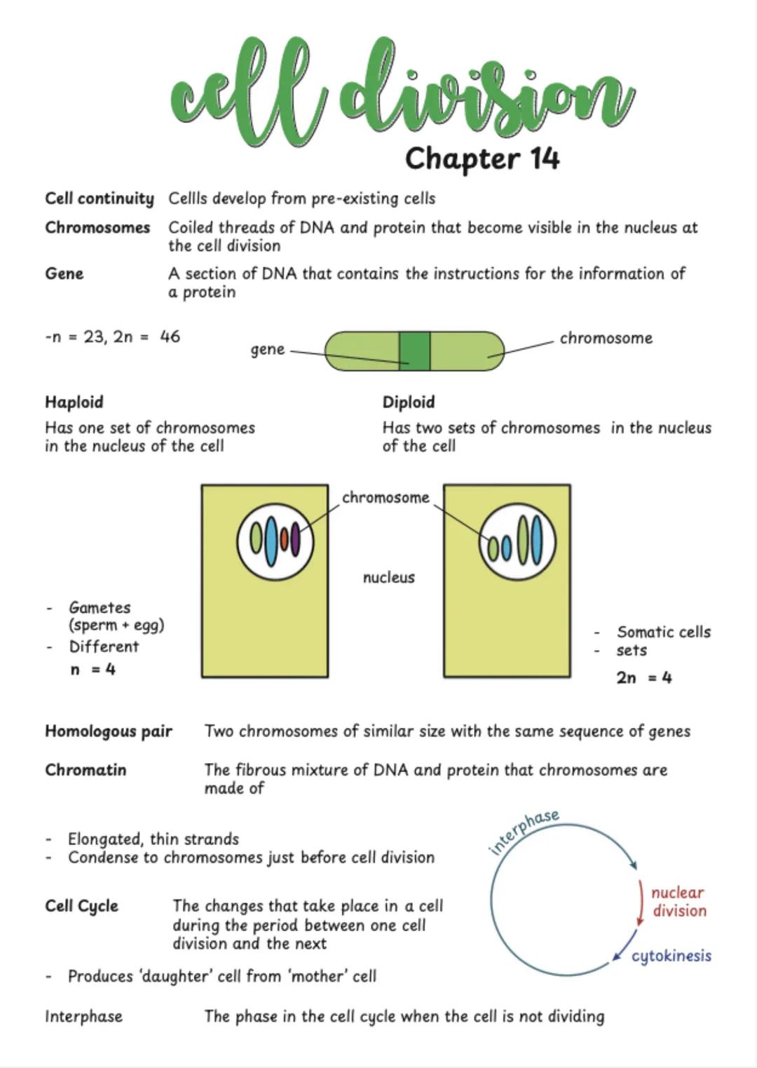
Cell Division Introduction
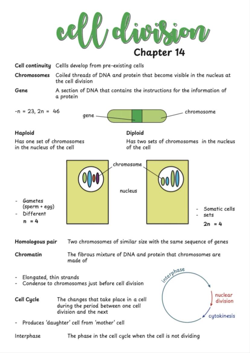
Cell continuity means all cells come from pre-existing cells - they don't just appear from nowhere. Understanding chromosomes (coiled DNA and protein threads) is essential since they carry genes that control protein production.
Diploid cells (like most of your body cells) have two sets of chromosomes . Haploid cells (sperm and egg) have just one set . When fertilisation happens, you get back to the full diploid number.
The cell cycle describes everything that happens between one cell division and the next. Most of this time is spent in interphase, when the cell grows, makes new organelles, and crucially, replicates its DNA ready for division.
Key Concept: Homologous pairs are matching chromosomes with the same genes - you get one from each parent!
6
of 10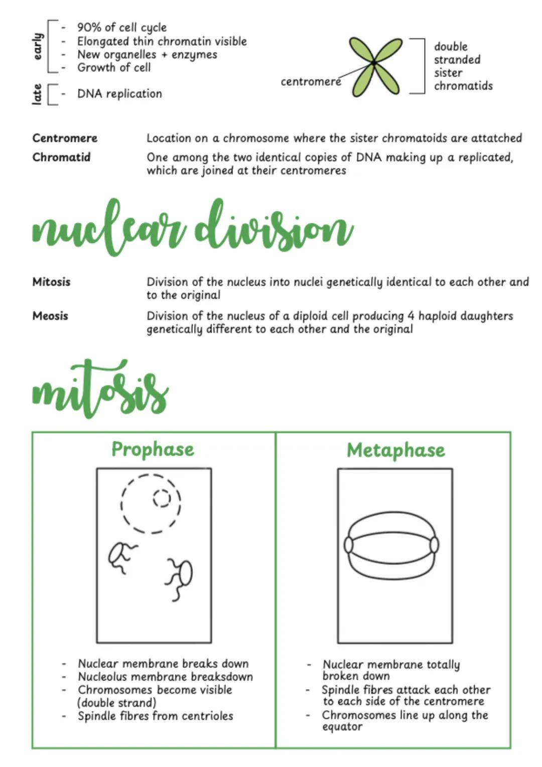
Mitosis Process
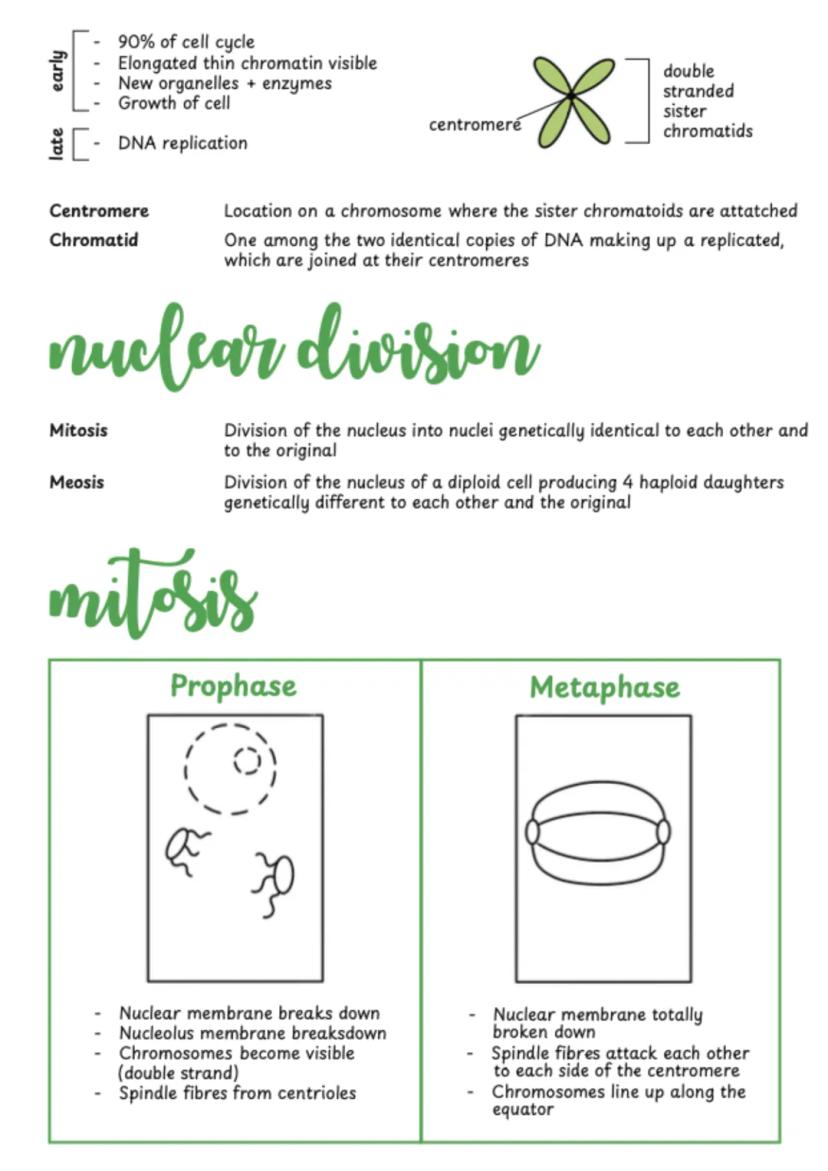
Interphase takes up 90% of the cell cycle. During this time, chromatin is elongated and thin, the cell grows, and DNA replication occurs. After replication, each chromosome consists of two identical sister chromatids joined at the centromere.
Mitosis has four clear stages you need to know. Prophase sees chromosomes becoming visible and the nuclear membrane breaking down. Metaphase is when chromosomes line up at the cell's equator, attached to spindle fibres.
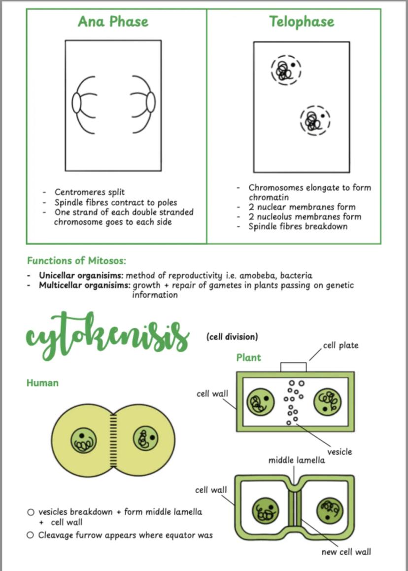
Anaphase is dramatic - centromeres split and sister chromatids are pulled to opposite poles by contracting spindle fibres. Telophase reverses the early changes: new nuclear membranes form and chromosomes elongate back to chromatin.
Memory Trick: Remember PMAT - Prophase, Metaphase, Anaphase, Telophase - for the order of mitotic stages!
7
of 10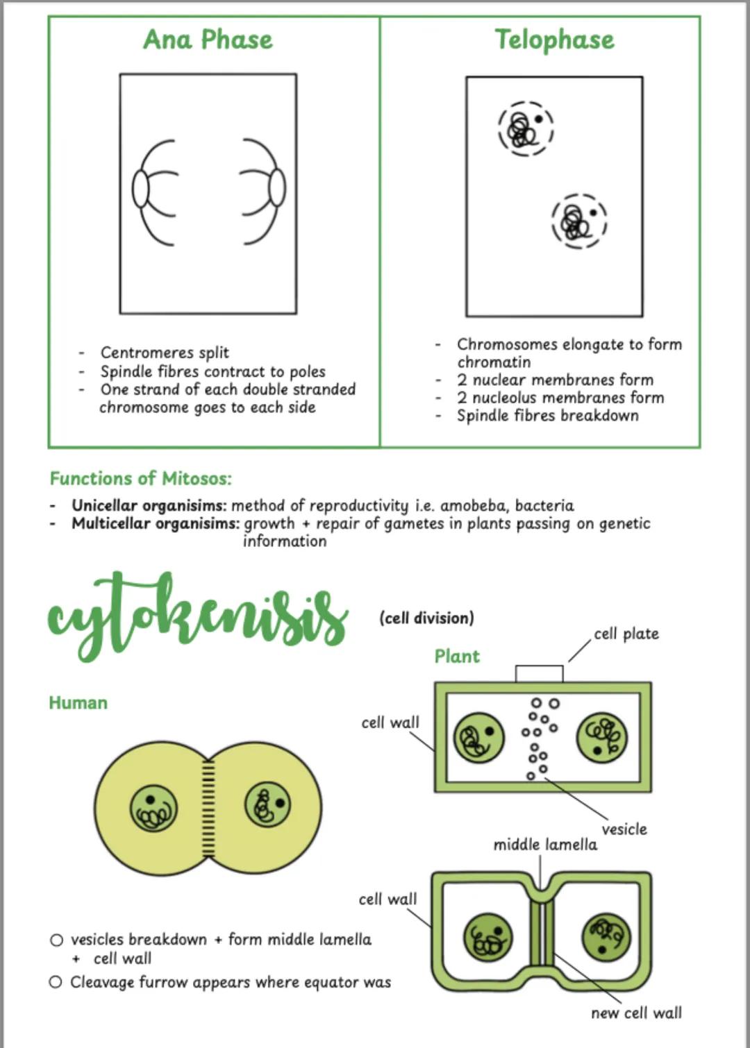
Cell Division Functions
Cytokinesis (actual cell splitting) happens differently in plants and animals. Animal cells form a cleavage furrow that pinches the cell in two. Plant cells can't pinch due to their rigid cell walls, so they build a new cell plate from the centre outwards.
Mitosis serves different purposes depending on the organism. In single-celled organisms like bacteria, it's their method of reproduction. In multicellular organisms like you, it's essential for growth and repair of damaged tissues.
The process ensures each new cell gets an identical copy of genetic information. This is crucial for maintaining consistent cell function throughout your body.
Real-world Application: Every time you heal from a cut, mitosis is working to replace damaged skin cells with identical healthy ones!
8
of 10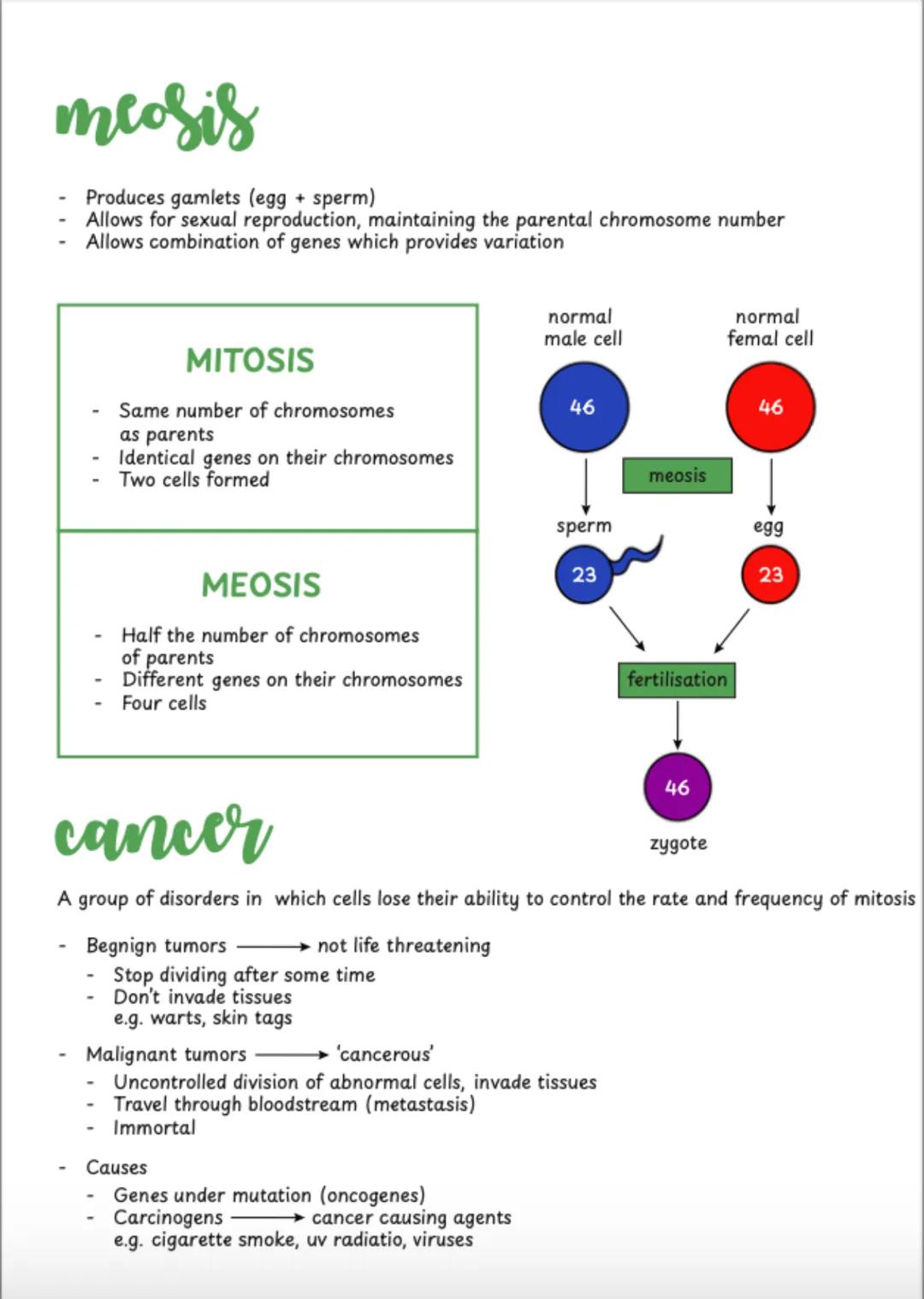
Meiosis and Cancer
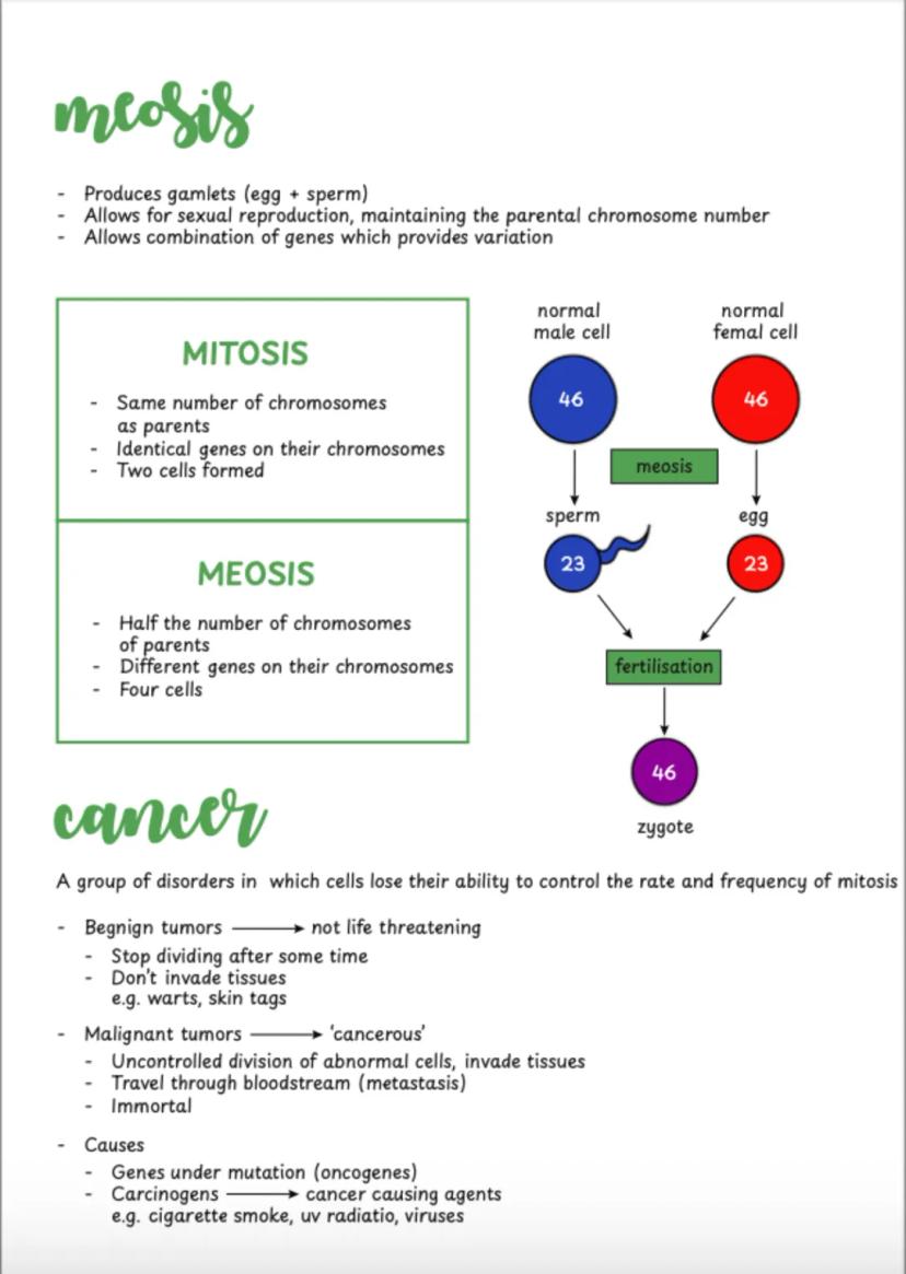
Meiosis produces gametes (sex cells) with half the chromosome number of parent cells. Unlike mitosis, meiosis creates genetic variation through gene combination, which is essential for evolution and species survival.
Sexual reproduction maintains species chromosome numbers across generations. When sperm meets egg , fertilisation produces a zygote with the full diploid number .
Cancer occurs when cells lose control over mitosis rates. Benign tumours eventually stop dividing and don't spread. Malignant tumours divide uncontrollably, invade other tissues, and can spread through the bloodstream (metastasis). Carcinogens like UV radiation and cigarette smoke damage genes that normally control cell division.
Health Connection: Understanding how normal cell division works helps explain why cancer treatments often target rapidly dividing cells!
9
of 10
Cell Organisation
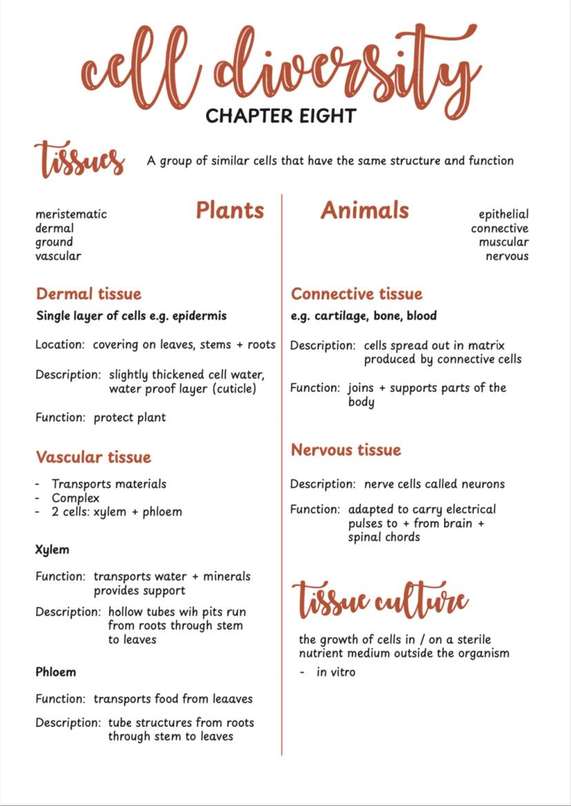
Tissues are groups of similar cells working together for the same function. Plants have four main tissue types: meristematic (growth), dermal (protection), ground (photosynthesis and storage), and vascular (transport).
Dermal tissue like epidermis protects plants with a waxy cuticle layer. Vascular tissue includes xylem (transports water and minerals upwards) and phloem (transports food from leaves to other parts).
Animal tissues include epithelial (covering surfaces), connective (support and joining), muscular (movement), and nervous (communication). Nervous tissue contains neurons that carry electrical signals around your body.
Study Strategy: Learn tissue types by their functions first, then worry about detailed structures - function determines form!
10
of 10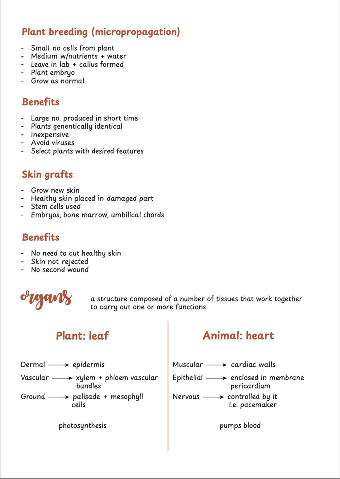
Tissue Culture and Organs
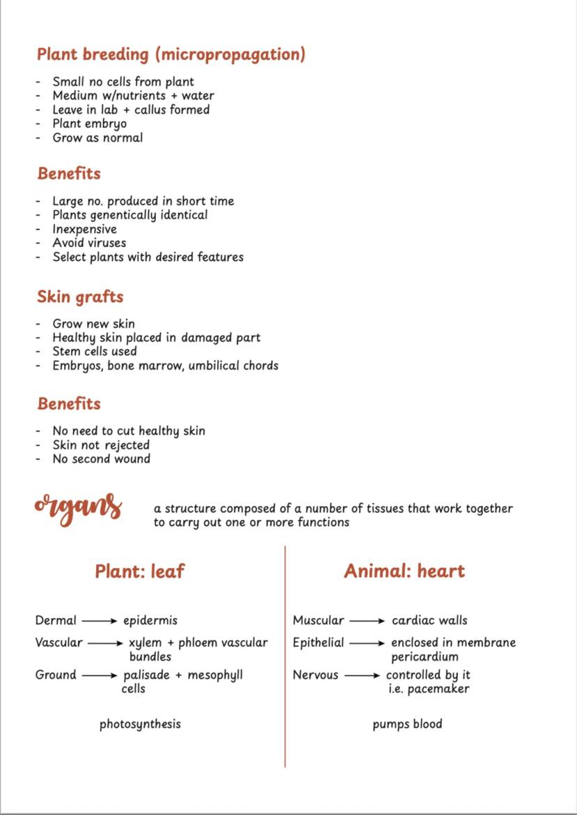
Tissue culture grows cells outside the organism in sterile conditions. Micropropagation in plants can produce thousands of identical plants from just a few cells, perfect for agriculture and conservation.
Medical applications include growing skin for grafts using stem cells from embryos, bone marrow, or umbilical cords. This avoids rejection issues and eliminates the need to damage healthy skin for traditional grafts.
Organs combine multiple tissues to perform complex functions. A leaf combines dermal tissue (epidermis), vascular tissue (transport), and ground tissue (photosynthesis). Your heart combines muscular tissue (pumping), epithelial tissue (lining), nervous tissue (pacemaker control), and connective tissue (support).
Future Focus: Tissue culture technology is revolutionising medicine and agriculture - understanding these basics opens doors to exciting career possibilities!
We thought you’d never ask...
What is the Knowunity AI companion?
Our AI companion is specifically built for the needs of students. Based on the millions of content pieces we have on the platform we can provide truly meaningful and relevant answers to students. But its not only about answers, the companion is even more about guiding students through their daily learning challenges, with personalised study plans, quizzes or content pieces in the chat and 100% personalisation based on the students skills and developments.
Where can I download the Knowunity app?
You can download the app in the Google Play Store and in the Apple App Store.
Is Knowunity really free of charge?
That's right! Enjoy free access to study content, connect with fellow students, and get instant help – all at your fingertips.
Most popular content in Biology
8Ecology introduction notes!
Start of the leaving cert ecology chapter
6th Year131
DNA & RNA
All notes on DNA & RNA including protein synthesis which is a HL topic
5th Year250
Vertebrates and Invertebrates
Students will distinguish between animals that have a backbone (vertebrates) and those that do not (invertebrates), identifying examples of each.
2nd Year240
Biomolecules: chapter 8
Summary and easily understandable notes to revise chapter 8 biomolecules. Includes good labelled diagrams for visual learners
5th Year210
Circulatory System
Students will learn about the heart, blood, and blood vessels, and how this system transports oxygen, nutrients, and waste products around the body.
3rd Year140
Respiration
All respiration notes including simple diagrams and glycolysis and the Krebs cycle
5th Year120
Photosynthesis : Biology
Photosynthesis
6th Year371
Plant Cells
Learning about the unique structures found in plant cells, such as the cell wall, chloroplasts, and large vacuole, and how they differ from animal cells.
1st Year80
Most popular content
9Irish oral questions and answers
Questions and answers for the leaving cert oral
5th Year4264
Key Quotes : Sive
Key Quotes and explanations: Sive
6th Year2842
Irish oral questions
Outline of oral questions
5th Year2055
Iníon- le hÁine Durkin
Aine Durkin’s poem, Iníon: Themes & summary
5th Year850
Irish poetry 2027
Iníon + Dínit an Bhróin
5th Year1133
LC HL notes- Iníon (poem)
Includes poem in English and Irish, theme, key words & phrases
5th Year2284
Cultural Context : Shawshank Redemption : Sive : Small Things Like These
Comparative Study : Cultural Context : Shawshank Redemption, Sive and Small Things Like These
6th Year1430
Mo Ghrá-sa (Idir Lúibíní)
Notes on mo ghrá-sa
5th Year380
An Gaeilge Aiste
Irish Language essay
6th Year1320
Can't find what you're looking for? Explore other subjects.
Students love us — and so will you.
4.6/5App Store
4.7/5Google Play
The app is very easy to use and well designed. I have found everything I was looking for so far and have been able to learn a lot from the presentations! I will definitely use the app for a class assignment! And of course it also helps a lot as an inspiration.
Stefan SiOS user
This app is really great. There are so many study notes and help [...]. My problem subject is French, for example, and the app has so many options for help. Thanks to this app, I have improved my French. I would recommend it to anyone.
Samantha KlichAndroid user
Wow, I am really amazed. I just tried the app because I've seen it advertised many times and was absolutely stunned. This app is THE HELP you want for school and above all, it offers so many things, such as workouts and fact sheets, which have been VERY helpful to me personally.
AnnaiOS user
Comprehensive LC Biology Notes and Diagrams
C
Chloe Ahern@chloeahern
Ever wondered what's actually inside the cells that make up your body? Cell structure and division are fundamental to understanding how all living things work, from the tiniest bacteria to complex organisms like humans and plants.
1
of 10
Sign up to see the content. It's free!
- Access to all documents
- Improve your grades
- Join milions of students
Cell Structure Basics
Your body is made up of trillions of cells, and understanding their ultrastructure (what you see under powerful microscopes) is key to biology success. Think of cells as tiny factories with different departments doing specific jobs.
Animal cells and plant cells share many features but have crucial differences. Both have a nucleus (the control centre), cytoplasm , and ribosomes (protein makers). The cell membrane acts like a selective bouncer, controlling what gets in and out using its phospholipid bilayer structure.
Mitochondria are your cell's power stations, producing energy through aerobic respiration. They're packed with folded membranes to maximise surface area - you'll find loads of them in active cells like neurons that need tons of energy.
Quick Tip: Remember that mitochondria have their own DNA and reproduce independently - they're like tiny organisms living inside your cells!
2
of 10Sign up to see the content. It's free!
- Access to all documents
- Improve your grades
- Join milions of students
Plant vs Animal Cells
Plant cells have some extra features that animal cells don't. The cell wall made of cellulose gives plants their rigid structure and shape - imagine it as a protective outer shell around the cell membrane.
Chloroplasts are the green powerhouses where photosynthesis happens. These double-membraned organelles contain chlorophyll and have stacked membranes called grana that capture sunlight. Plant cells also have a large central vacuole filled with sap that helps maintain the plant's shape and stores materials.
The nucleus is the brain of any cell, containing chromosomes (which become chromatin when not dividing). Nuclear pores let important molecules like mRNA pass through. Cytoplasm is 90% water and houses all the organelles where metabolic reactions occur.
Remember: All the living parts of a cell together are called protoplasm - this includes everything except the cell wall!
3
of 10Sign up to see the content. It's free!
- Access to all documents
- Improve your grades
- Join milions of students
Prokaryotic vs Eukaryotic Cells
Understanding the difference between eukaryotic and prokaryotic cells is crucial for your exams. Eukaryotic cells (like yours) have a membrane-bound nucleus and organelles - they're the advanced, complex cells found in animals, plants, and fungi.
Prokaryotic cells are much simpler - think bacteria. They have no membrane-bound nucleus, just DNA floating in a circular loop. No fancy organelles either, and they're much smaller than eukaryotic cells.
Light microscopes can magnify up to 1,500 times and show basic cell structures. Electron microscopes are the real game-changers, magnifying up to 1,000,000 times and revealing incredible detail. Understanding how to use a light microscope properly is essential lab skill.
Lab Success: Always start with low power objective lens, use coarse focus first, then switch to high power and use fine focus for crisp images!
4
of 10Sign up to see the content. It's free!
- Access to all documents
- Improve your grades
- Join milions of students
Microscope Experiments
Preparing cell samples is a key practical skill you'll need to master. For plant cells, onion epidermis works brilliantly - it's thin, easy to handle, and shows cell walls clearly when stained with iodine solution.
Animal cells from your cheek are perfect for observation. A gentle rub with a cotton bud gives you plenty of cells to examine. Methylene blue stain makes the nucleus and cell membrane visible under the microscope.
The technique is crucial: place your sample on a slide with a drop of water, add stain, then lower the coverslip at an angle to avoid air bubbles. Always blot excess liquid to prevent mess and improve image quality.
Pro Tip: When switching from low to high power, always centre your specimen first - high power has a much smaller field of view!
5
of 10Sign up to see the content. It's free!
- Access to all documents
- Improve your grades
- Join milions of students
Cell Division Introduction
Cell continuity means all cells come from pre-existing cells - they don't just appear from nowhere. Understanding chromosomes (coiled DNA and protein threads) is essential since they carry genes that control protein production.
Diploid cells (like most of your body cells) have two sets of chromosomes . Haploid cells (sperm and egg) have just one set . When fertilisation happens, you get back to the full diploid number.
The cell cycle describes everything that happens between one cell division and the next. Most of this time is spent in interphase, when the cell grows, makes new organelles, and crucially, replicates its DNA ready for division.
Key Concept: Homologous pairs are matching chromosomes with the same genes - you get one from each parent!
6
of 10Sign up to see the content. It's free!
- Access to all documents
- Improve your grades
- Join milions of students
Mitosis Process
Interphase takes up 90% of the cell cycle. During this time, chromatin is elongated and thin, the cell grows, and DNA replication occurs. After replication, each chromosome consists of two identical sister chromatids joined at the centromere.
Mitosis has four clear stages you need to know. Prophase sees chromosomes becoming visible and the nuclear membrane breaking down. Metaphase is when chromosomes line up at the cell's equator, attached to spindle fibres.
Anaphase is dramatic - centromeres split and sister chromatids are pulled to opposite poles by contracting spindle fibres. Telophase reverses the early changes: new nuclear membranes form and chromosomes elongate back to chromatin.
Memory Trick: Remember PMAT - Prophase, Metaphase, Anaphase, Telophase - for the order of mitotic stages!
7
of 10Sign up to see the content. It's free!
- Access to all documents
- Improve your grades
- Join milions of students
Cell Division Functions
Cytokinesis (actual cell splitting) happens differently in plants and animals. Animal cells form a cleavage furrow that pinches the cell in two. Plant cells can't pinch due to their rigid cell walls, so they build a new cell plate from the centre outwards.
Mitosis serves different purposes depending on the organism. In single-celled organisms like bacteria, it's their method of reproduction. In multicellular organisms like you, it's essential for growth and repair of damaged tissues.
The process ensures each new cell gets an identical copy of genetic information. This is crucial for maintaining consistent cell function throughout your body.
Real-world Application: Every time you heal from a cut, mitosis is working to replace damaged skin cells with identical healthy ones!
8
of 10Sign up to see the content. It's free!
- Access to all documents
- Improve your grades
- Join milions of students
Meiosis and Cancer
Meiosis produces gametes (sex cells) with half the chromosome number of parent cells. Unlike mitosis, meiosis creates genetic variation through gene combination, which is essential for evolution and species survival.
Sexual reproduction maintains species chromosome numbers across generations. When sperm meets egg , fertilisation produces a zygote with the full diploid number .
Cancer occurs when cells lose control over mitosis rates. Benign tumours eventually stop dividing and don't spread. Malignant tumours divide uncontrollably, invade other tissues, and can spread through the bloodstream (metastasis). Carcinogens like UV radiation and cigarette smoke damage genes that normally control cell division.
Health Connection: Understanding how normal cell division works helps explain why cancer treatments often target rapidly dividing cells!
9
of 10Sign up to see the content. It's free!
- Access to all documents
- Improve your grades
- Join milions of students
Cell Organisation
Tissues are groups of similar cells working together for the same function. Plants have four main tissue types: meristematic (growth), dermal (protection), ground (photosynthesis and storage), and vascular (transport).
Dermal tissue like epidermis protects plants with a waxy cuticle layer. Vascular tissue includes xylem (transports water and minerals upwards) and phloem (transports food from leaves to other parts).
Animal tissues include epithelial (covering surfaces), connective (support and joining), muscular (movement), and nervous (communication). Nervous tissue contains neurons that carry electrical signals around your body.
Study Strategy: Learn tissue types by their functions first, then worry about detailed structures - function determines form!
10
of 10Sign up to see the content. It's free!
- Access to all documents
- Improve your grades
- Join milions of students
Tissue Culture and Organs
Tissue culture grows cells outside the organism in sterile conditions. Micropropagation in plants can produce thousands of identical plants from just a few cells, perfect for agriculture and conservation.
Medical applications include growing skin for grafts using stem cells from embryos, bone marrow, or umbilical cords. This avoids rejection issues and eliminates the need to damage healthy skin for traditional grafts.
Organs combine multiple tissues to perform complex functions. A leaf combines dermal tissue (epidermis), vascular tissue (transport), and ground tissue (photosynthesis). Your heart combines muscular tissue (pumping), epithelial tissue (lining), nervous tissue (pacemaker control), and connective tissue (support).
Future Focus: Tissue culture technology is revolutionising medicine and agriculture - understanding these basics opens doors to exciting career possibilities!
We thought you’d never ask...
What is the Knowunity AI companion?
Our AI companion is specifically built for the needs of students. Based on the millions of content pieces we have on the platform we can provide truly meaningful and relevant answers to students. But its not only about answers, the companion is even more about guiding students through their daily learning challenges, with personalised study plans, quizzes or content pieces in the chat and 100% personalisation based on the students skills and developments.
Where can I download the Knowunity app?
You can download the app in the Google Play Store and in the Apple App Store.
Is Knowunity really free of charge?
That's right! Enjoy free access to study content, connect with fellow students, and get instant help – all at your fingertips.
Most popular content in Biology
8Ecology introduction notes!
Start of the leaving cert ecology chapter
6th Year131
DNA & RNA
All notes on DNA & RNA including protein synthesis which is a HL topic
5th Year250
Vertebrates and Invertebrates
Students will distinguish between animals that have a backbone (vertebrates) and those that do not (invertebrates), identifying examples of each.
2nd Year240
Biomolecules: chapter 8
Summary and easily understandable notes to revise chapter 8 biomolecules. Includes good labelled diagrams for visual learners
5th Year210
Circulatory System
Students will learn about the heart, blood, and blood vessels, and how this system transports oxygen, nutrients, and waste products around the body.
3rd Year140
Respiration
All respiration notes including simple diagrams and glycolysis and the Krebs cycle
5th Year120
Photosynthesis : Biology
Photosynthesis
6th Year371
Plant Cells
Learning about the unique structures found in plant cells, such as the cell wall, chloroplasts, and large vacuole, and how they differ from animal cells.
1st Year80
Most popular content
9Irish oral questions and answers
Questions and answers for the leaving cert oral
5th Year4264
Key Quotes : Sive
Key Quotes and explanations: Sive
6th Year2842
Irish oral questions
Outline of oral questions
5th Year2055
Iníon- le hÁine Durkin
Aine Durkin’s poem, Iníon: Themes & summary
5th Year850
Irish poetry 2027
Iníon + Dínit an Bhróin
5th Year1133
LC HL notes- Iníon (poem)
Includes poem in English and Irish, theme, key words & phrases
5th Year2284
Cultural Context : Shawshank Redemption : Sive : Small Things Like These
Comparative Study : Cultural Context : Shawshank Redemption, Sive and Small Things Like These
6th Year1430
Mo Ghrá-sa (Idir Lúibíní)
Notes on mo ghrá-sa
5th Year380
An Gaeilge Aiste
Irish Language essay
6th Year1320
Can't find what you're looking for? Explore other subjects.
Students love us — and so will you.
4.6/5App Store
4.7/5Google Play
The app is very easy to use and well designed. I have found everything I was looking for so far and have been able to learn a lot from the presentations! I will definitely use the app for a class assignment! And of course it also helps a lot as an inspiration.
Stefan SiOS user
This app is really great. There are so many study notes and help [...]. My problem subject is French, for example, and the app has so many options for help. Thanks to this app, I have improved my French. I would recommend it to anyone.
Samantha KlichAndroid user
Wow, I am really amazed. I just tried the app because I've seen it advertised many times and was absolutely stunned. This app is THE HELP you want for school and above all, it offers so many things, such as workouts and fact sheets, which have been VERY helpful to me personally.
AnnaiOS user